

Author: Vigor Forty Editorial Team
Medically Reviewed By: Editorial Review Board
Disclaimer: This article is for informational purposes only and is not a substitute for professional medical advice. Read our full Medical Disclaimer.
📌 Key Takeaways
- Sciatica and Piriformis Syndrome Are Not the Same Thing—But They Feel Identical: Both cause pain, tingling, or numbness that radiates down the back of the leg. However, the underlying cause is fundamentally different—and treating the wrong condition can worsen symptoms.
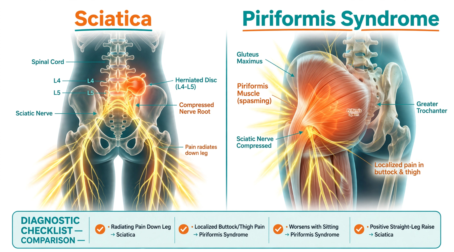
- Sciatica Is a Nerve Root Problem (Spine): Compression or irritation of the sciatic nerve roots (L4–S3) in the lumbar spine. Causes include herniated discs, bone spurs, or spinal stenosis. The pain often follows a specific dermatomal pattern (e.g., down the back of the thigh to the calf).
- Piriformis Syndrome Is a Muscle Problem (Buttock): The piriformis muscle in the deep gluteal region spasms and compresses the sciatic nerve as it passes through or under the muscle. The pain is typically localized to the buttock and may radiate down the leg, but it rarely goes below the knee.
- The “Sitting Test” Is the Best Home Diagnostic: If your pain worsens when sitting on a hard surface (and improves when standing/walking), it’s likely piriformis syndrome. If your pain worsens when bending forward (flexion) or coughing/sneezing, it’s likely sciatica from a disc issue.
- The Treatment Protocols Are Completely Different: Sciatica responds to spinal extension (McKenzie press-ups) and avoiding flexion. Piriformis syndrome responds to deep gluteal stretching (Figure-4), massage, and strengthening the surrounding hip muscles. Mixing them up is a recipe for frustration.
Introduction: The Pain That Makes No Sense
You feel it deep in your buttock—a dull ache that sometimes shoots down the back of your thigh. Sometimes it tingles. Sometimes it burns. You google your symptoms, and the internet gives you the same confusing answer: “Sciatica.” You start doing stretches for sciatica—lying on your back, pulling your knee to your chest, twisting your spine. But the pain gets worse. Much worse.
What you are experiencing might be sciatica. Or it might be piriformis syndrome. The symptoms are nearly identical. The mechanisms are completely different. And the treatments are opposite.
Sciatica is a neurological condition. The sciatic nerve roots in your lower spine are being pinched by a disc bulge, bone spur, or arthritic narrowing. The nerve is literally being compressed at its source.
Piriformis Syndrome is a muscular condition. The sciatic nerve runs through or under the piriformis muscle—a deep rotator of your hip. When this muscle spasms, tightens, or becomes inflamed, it compresses the nerve after it has left the spine.
The distinction matters. If you treat a disc herniation (sciatica) with aggressive gluteal stretching (piriformis treatment), you might provoke more nerve root irritation. If you treat a spasming piriformis muscle with spinal extension exercises (sciatica treatment), you are ignoring the root cause.
This article gives you the definitive guide to telling these two conditions apart—using simple home tests—and provides targeted treatment protocols for each. By the end, you will know exactly which condition you have and what to do about it.
Section 1: What Is Sciatica?
The Definition:
Sciatica is not a diagnosis—it is a symptom. It refers to pain, weakness, numbness, or tingling that radiates along the path of the sciatic nerve, which runs from your lower spine, through your buttock, and down the back of your leg to your foot.
The Mechanism:
The sciatic nerve is formed by the nerve roots of L4, L5, S1, S2, and S3. These roots exit the spine through the intervertebral foramina (the holes between the vertebrae). When these roots are compressed or irritated, the nerve signals misfire, causing pain, paresthesia (tingling), and sometimes weakness.
Common Causes:
- Lumbar disc herniation (most common): The nucleus pulposus of the disc bulges backward and compresses a nerve root. L4-L5 and L5-S1 are the most frequently affected levels.
- Lumbar spinal stenosis: Narrowing of the spinal canal, usually from facet joint arthritis and ligamentum flavum hypertrophy. This compresses multiple nerve roots, causing bilateral symptoms and neurogenic claudication (pain when walking).
- Spondylolisthesis: A vertebral body slips forward over the one below it, narrowing the foramen.
- Tumors or infections (rare): Mass lesions compressing the nerve roots.
Symptoms:
- Sharp, shooting, or burning pain that originates in the lower back or buttock and radiates down the leg.
- Pain often follows a dermatomal pattern: L4 radiates to the front of the thigh; L5 radiates down the lateral (outside) of the calf to the top of the foot; S1 radiates down the back of the calf to the heel and lateral foot.
- Numbness or tingling in the same distribution.
- Weakness in the affected leg, foot drop (difficulty lifting the front of the foot), or loss of reflex (ankle jerk).
- Symptoms often worsen with spinal flexion (bending forward, sitting slouched), coughing, sneezing, or straining (Valsalva maneuver).
Section 2: What Is Piriformis Syndrome?
The Definition:
Piriformis syndrome is a neuromuscular disorder characterized by spasms of the piriformis muscle, which then compress the sciatic nerve as it passes through the greater sciatic notch.
The Anatomy:
The piriformis muscle originates from the anterior surface of the sacrum, passes through the greater sciatic foramen, and attaches to the greater trochanter of the femur. It is a lateral rotator of the hip (turns the leg outward) and an abductor (moves the leg away from the body) when the hip is flexed.
The sciatic nerve typically passes under the piriformis. However, in approximately 15–20% of the population, the nerve passes through the piriformis muscle belly or splits around it. This anatomical variation makes those individuals significantly more susceptible to piriformis syndrome.
The Mechanism:
Prolonged sitting, trauma (a fall on the buttock), repetitive overuse (running, cycling), or muscle imbalances cause the piriformis to spasm or become hypertrophied (enlarged). The spasming muscle compresses the sciatic nerve, causing referred pain down the leg.
Symptoms:
- Deep, aching pain in the buttock (often unilateral).
- Pain that radiates down the back of the thigh, but rarely below the knee. (This is a key differentiator.)
- Pain that worsens with sitting—especially on hard surfaces—and improves with standing or walking.
- Pain that worsens with specific movements: hip adduction (crossing the leg) and internal rotation (turning the foot inward).
- Tenderness to palpation (deep pressure) over the piriformis muscle—located roughly in the middle of the buttock, about 2–3 inches lateral to the sacrum.
- Absence of true lower back pain (though the buttock pain can refer to the lower back).
Section 3: The Key Differences—At a Glance
| Feature | Sciatica (Nerve Root Compression) | Piriformis Syndrome (Muscle Compression) |
|---|---|---|
| Origin | Lumbar spine (L4–S3 nerve roots) | Piriformis muscle in the deep gluteal region |
| Pain Location | Lower back, buttock, radiating down the leg | Primarily deep gluteal (buttock), radiating to thigh |
| Pain Below Knee | Common (radiates to calf, foot, toes) | Rare (usually stops at or above the knee) |
| Worsened By | Spinal flexion (bending forward), coughing, sneezing, sitting slouched | Sitting on hard surfaces, hip adduction/internal rotation |
| Improved By | Standing, walking (if stenosis), spinal extension (leaning back) | Standing, walking, warm baths, massage |
| Neurological Signs | Weakness, numbness, reflex loss (positive straight leg raise) | Usually absent; muscle strength normal |
| Palpation | Tenderness over lumbar spine (paraspinal) | Tenderness over piriformis (deep buttock, 2–3 inches from sacrum) |
| Diagnostic Test | MRI of lumbar spine | MRI of pelvis (rare), diagnostic ultrasound, or injection of anesthetic into piriformis |
Section 4: The “Sitting Test”—The Simplest Home Diagnostic
This is the most practical test you can perform at home. It costs nothing and takes 30 seconds.
The Test:
- Sit on a hard, unpadded surface (like a wooden chair or the floor) for 30 seconds.
- Notice your pain level.
- Stand up and walk around for 30 seconds.
- Notice your pain level.
The Interpretation:
- If your pain significantly worsens when sitting and significantly improves when standing/walking → Likely Piriformis Syndrome. The piriformis is compressed against the hard surface, directly irritating the sciatic nerve. Walking relaxes the muscle.
- If your pain is relatively unchanged by sitting and is worse with bending forward or coughing → Likely Sciatica. Flexion increases intradiscal pressure, pushing the disc against the nerve root. Standing may not dramatically change it.
The Caveat:
Some individuals have both conditions—a disc herniation that also triggers compensatory piriformis spasm. This is called “double crush” syndrome. In this case, you will need to treat both.
Section 5: Treatment Protocol for Sciatica
If your pain is consistent with nerve root compression, the primary goal is to decompress the nerve by reducing intradiscal pressure and avoiding flexion.
Phase 1: Acute (First 48 Hours)
- Avoid spinal flexion. Do not bend forward. Do not do toe-touches. Do not pull your knees to your chest. Do not sit slouched.
- Perform McKenzie Press-Ups (Prone Extension): Lie on your stomach, place your forearms on the floor, and gently press up, straightening your elbows. Your lower back should sag into extension. This encourages the disc nucleus to migrate anteriorly, away from the nerve root. Hold for 5 seconds. Repeat 10 times every 2 hours.
- Ice, not heat: Apply an ice pack to the lower back (20 minutes on, 20 minutes off) to reduce inflammation. Heat can increase swelling.
- Consider NSAIDs (ibuprofen, naproxen) for 3–5 days if you have no contraindications.
- Avoid heavy lifting and high-impact activities.
Phase 2: Subacute (1–4 Weeks)
- Continue McKenzie Press-Ups as a daily maintenance routine.
- Add side-glides (lateral shifts) if the pain is asymmetrical (e.g., pain is worse on one side). This mobilizes the lumbar spine in the transverse plane.
- Begin gentle core stabilization: Dead bug exercises (lying on your back, alternating arm/leg reaches) without flexing the spine. Keep your back flat on the floor.
- Walking: Begin with 5–10 minutes of walking daily. Increase gradually. Walking is one of the best activities for discogenic pain—it promotes disc hydration through natural pumping.
- Avoid sitting for >45 minutes at a time. Stand up and walk around.
Phase 3: Long-Term (4+ Weeks)
- Strengthen the core and glutes: Bird-dogs, planks (modified), and glute bridges (with posterior pelvic tilt).
- Address ergonomics: Ensure your desk chair has adequate lumbar support (see Article #5).
- Consider consulting a physical therapist for a tailored extension-based program.
- If symptoms persist beyond 6 weeks, or if you develop progressive weakness, bowel/bladder changes, or saddle anesthesia (numbness in the groin), seek immediate medical attention—these are “cauda equina” red flags requiring emergency evaluation.
Section 6: Treatment Protocol for Piriformis Syndrome
If your pain is consistent with piriformis compression, the primary goal is to relax the spasming muscle and address the underlying biomechanical imbalances.
Phase 1: Acute (First 48 Hours)
- Deep gluteal massage: Use a tennis ball, lacrosse ball, or massage gun to apply deep, sustained pressure to the piriformis. Locate the tender spot in the middle of the buttock (about 2–3 inches lateral to the sacrum). Press into it with your body weight for 30–60 seconds. You should feel a deep, releasing ache—not sharp nerve pain.
- Heat, not ice: Apply a heating pad to the buttock to relax the muscle spasm. Ice can increase spasm in muscle tissue.
- Perform the Seated Figure-4 Stretch: Sit on a chair with a tall spine. Cross your right ankle over your left knee. Gently lean forward from the hips (keeping your back straight) until you feel a stretch in the right glute. Hold for 30 seconds. Repeat 3 times per side. (See Article #3 for details.)
- Avoid sitting on hard surfaces. Use a padded cushion or stand for part of your day.
Phase 2: Subacute (1–4 Weeks)
- Strengthen the surrounding hip muscles: Glute medius weakness is a primary driver of piriformis overcompensation. Perform Lateral Band Walks (Article #4) 3x per week.
- Add pigeon pose (modified): Lie on your back with your right ankle crossed over your left knee (Figure-4). Gently pull your left knee toward your chest until you feel a stretch in the right glute. This is a gentler version of the full pigeon pose, avoiding lumbar flexion.
- Eccentric hamstring strengthening: Strong hamstrings offload the piriformis (which assists in hip extension). Perform Romanian Deadlifts (RDLs) with a light weight (Article #2).
- Stretch the hip flexors: Tight hip flexors pull the pelvis into anterior tilt, which increases the stretch on the piriformis. Perform kneeling hip flexor stretches.
Phase 3: Long-Term (4+ Weeks)
- Address sitting habits: If you sit for long hours, use a cushion with a cutout (e.g., a “seat cushion for coccyx” or “donut cushion”) that offloads the piriformis.
- Regular stretching routine: Incorporate Figure-4 stretches into your daily morning routine (Article #6).
- Consider trigger point injections: If conservative measures fail, a physician can perform a guided injection of anesthetic and corticosteroid directly into the piriformis. This often provides rapid, significant relief.
- Review running/cycling form: If you are an endurance athlete, consider a gait analysis to identify muscular imbalances.
Section 7: The Straight Leg Raise (SLR) Test—A Quick Nerve Screen
This simple test helps differentiate between nerve root irritation (sciatica) and muscle tightness (piriformis syndrome). It should be performed gently and only to the point of first discomfort.
How:
- Lie on your back with your legs straight.
- Slowly raise the affected leg straight up (with your knee locked) by lifting it off the floor. Keep your opposite leg flat.
- Note the angle at which you first feel pain (not just stretch).
- Perform the same test on the unaffected leg.
Interpretation:
- If you feel pain in the affected leg at <60 degrees of elevation, and this pain is worse than the unaffected leg, it suggests nerve root irritation (sciatica) . The sciatic nerve is being pulled and compressed at the spinal level.
- If you feel a similar stretch/tightness in both legs at >70 degrees, it suggests muscle tightness (hamstrings or piriformis) , not nerve root compression.
Important: If the test reproduces your leg pain, stop immediately. Do not push through pain. This is a screening tool, not a treatment.
Section 8: FAQ—Your Top Sciatica and Piriformis Questions
Q: Can I have both sciatica and piriformis syndrome at the same time?
A: Yes. This is called the “double crush syndrome.” A lumbar disc herniation can irritate the nerve root, and the pain can cause compensatory piriformis spasm, which further compresses the nerve. In this case, treat both: McKenzie press-ups for the spine + Figure-4 stretches and massage for the piriformis.
Q: When should I see a doctor for leg pain?
A: Seek immediate medical attention if you experience:
- Progressive weakness (foot drop, cannot stand on toes or heels).
- Loss of bowel or bladder control.
- Saddle anesthesia (numbness in the groin, perineum, or inner thighs).
These are red flags for cauda equina syndrome, a surgical emergency.
For non-emergency symptoms, see a doctor if conservative measures fail after 4–6 weeks.
Q: Can a chiropractor help with these conditions?
A: Yes, for sciatica. Spinal manipulation and mobilization can reduce disc pressure and improve segmental motion. For piriformis syndrome, chiropractors may use soft tissue therapy (ART—Active Release Technique) and mobilization of the sacroiliac joint. Choose a chiropractor with a sports medicine or orthopaedic focus.
Q: I’ve been doing the McKenzie press-ups, but my buttock pain is worse. What does that mean?
A: It suggests your pain is likely piriformis syndrome, not sciatica. The press-ups are extending your spine and potentially increasing the stretch on the already-spasming piriformis. Stop press-ups and switch to deep gluteal massage and Figure-4 stretches.
Q: Is surgery ever necessary for these conditions?
A: For sciatica, surgery (microdiscectomy) is indicated if you have progressive neurological deficits or if 6–8 weeks of conservative care fails. For piriformis syndrome, surgery (piriformis release) is exceedingly rare and only considered after all other treatments have failed. Both are last-resort options.
Q: Can exercise make piriformis syndrome worse?
A: Yes. Activities that involve repetitive hip adduction and internal rotation—like cycling, rowing, or running on a banked track—can aggravate it. Avoid these during the acute phase. Focus on glute strengthening and stretching instead.
The VigorForty Take
Pain in your buttock and leg is frustrating. It limits your mobility, disrupts your sleep, and can make you feel like you are aging faster than you actually are. But the first step to relief is accurate diagnosis.
The distinction between sciatica (nerve root compression) and piriformis syndrome (muscle compression) is not an academic exercise—it is the difference between doing the right stretches and doing the wrong ones. Treating a disc herniation with aggressive gluteal stretching can worsen nerve root irritation. Treating a spasming piriformis with spinal extension can leave you frustrated and in pain.
Use the Sitting Test and the Straight Leg Raise as your initial diagnostic tools. If you are still unsure, see a physical therapist or sports medicine physician who can perform a comprehensive neurological and musculoskeletal exam. A targeted MRI or ultrasound may be needed to confirm the diagnosis.
Once you know what you are dealing with, the treatment protocols are clear: extension and core stabilization for sciatica; deep gluteal stretching, massage, and hip strengthening for piriformis syndrome.
Your Challenge: Spend 3 days following the diagnostic protocol. Sit on a hard surface, stand up, walk, and note the pattern. If it is sciatica, stop all forward bends immediately. If it is piriformis syndrome, grab a tennis ball and start releasing that muscle. Within a week, you will likely see significant improvement—and you will finally understand the pain that has been confusing you for months.
Read All Realated Articles
- The Complete Guide to Heart Health: Beyond Omega-3s—CoQ10, Magnesium, Potassium, and Nitric Oxide Support After 40
- Why Your Hamstrings Are Tight (And Stretching Them Won’t Fix It)—The Real Causes & 4 Fixes for Men Over 40
- The 3 Worst Stretches for the Over-40 Lower Back (And What to Do Instead)
- Pickleball and Your Knees: 4 Preventative Exercises to Bulletproof Your Joints (For Men Over 40)
- The Desk Chair Evaluation: How to Setup Your Home Office to Protect Your Neck, Shoulders, and Upper Back (For Men Over 40)
- The 5-Minute Morning Mobility Routine for Americans Over 40—Start Your Day Pain-Free
- Is Your Pillow Causing Your Neck Pain? The Over-40 Guide to Sleep Posture, Pillow Selection, and Morning Relief
- Collagen vs. Glucosamine vs. Omega-3s: The 3 Best Supplements for Joint Pain (2026 Update for Men Over 40)
- Sciatica vs. Piriformis Syndrome: How to Tell the Difference and Find the Right Treatment (For Men Over 40)
- The 3 Worst Exercises for Bad Knees (And What to Do Instead)—A 40+ Gym Survival Guide
- How to Improve Your Balance After 40—The Underrated Longevity Skill Every Man Needs